XPD/RESPES/RESPED
Resonant Photoemission Spectroscopy/Diffraction
Characterisation Installation 5
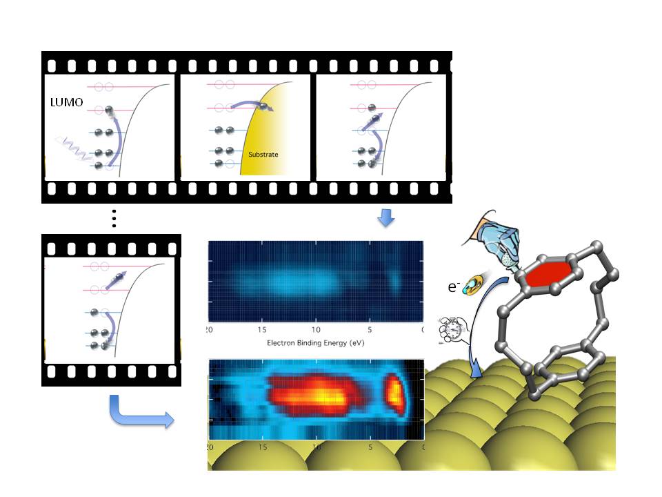
Photoelectron Diffraction (PED) is a structural technique that probes the local atomic arrangement around a chemically selected emitter. On the other hand Resonant photoemission (RESPES) is a spectroscopic technique that provides insight into the contributions of different atomic species to the electronic states in the valence/conduction region, thus allowing to attribute a line component to one or another element in a complex system.
The combination of these two techniques can provide information not only on the electronic properties, but also on the order of the desired components. In addition, the resonance condition enhances the intensity of the specific spectral line thus permitting the measurement of full angular PED patterns also on spectral features that would be normally too faint for a reliable detection with respect to the background of secondary electrons. The latter specific advantage can be exploited not only to enhance the valence band signal, whose photoemission cross-section drops quickly at the photon energy (hence electron kinetic energy) required for an useful PED study, but also to enhance the Auger transitions at the corresponding ionization edge, as may be required in the case of a low coverage of the selected atomic species, or in the case of searching specific electronic effects.
At variance with conventional PED, the photoemission at the resonance of selected ionization thresholds cannot be performed with a standard X-ray lamp, and the fine tuning of the photon energy provided by a Synchrotron radiation source is necessary.
ARPES, XPD - PEARL Beamline @ Swiss Light Source Synchrotron
The PEARL (Photoemission and atomic resolution laboratory) beamline is dedicated to the structural characterisation of local bonding geometry of molecular adsorbates on metal or semiconductor surfaces, of nanostructured surfaces, and of surfaces of complex materials. It is a soft X-ray beamline with an angle-resolved photoelectron spectrometer for angle-scanned and photon energy-scanned X-ray photoelectron diffraction (XPD)
XPD/RESPES/RESPED - BACH Beamline @ Elettra Synchrotron
Elettra synchrotron, Apple II undulators; variable polarization (horizontal, vertical, circular ±); beam size on the sample 350x350 (HxV, µm2), vertical size can be reduced on request, flux on sample @10 µm slits (best resolution) (ph./s) 2x10¹² - 6x10¹⁰; resolving power (E/ΔE): 20000 @44eV -5000 (@1650eV)
Many samples can be accommodated in a 30x30 mm2 area; sample size: 0.5 mm-28 mm; measurement: 40/50K- 500 K; preparation: 40/50K-1200 K
Heating and cooling (ebeam, direct current, PBN), ion gun (VG), 4 evaporator ports (CF40), gas inlet valve (variable leak valve), diamond file scraper, cleaver, LEED (OciLEED); evaporators for organic molecules; e-beam evaporator (Omicron) for metals (evaporation at low sample temperatures is also possible); load lock

