XAS
X-ray Absorption Spectroscopy
Characterisation Installation 5
XAS is a widely used technique for determining the local electronic, structural and magnetic properties of matter in different scientific fields including molecular and condensed matter physics, material science, engineering, chemistry, earth science and biology.
When the incident photon energy is scanned through the energy of a core shell, there is an abrupt increase in the absorption cross-section. This gives rise to a so-called absorption edge representing a particular core shell. These edges are at different energies due to the different nuclear charges of each element and different orbitals involved, making XAS an element and orbital selective technique.
XAS is very sensitive to the local bonding environment of the absorbing atom, providing information with regard to oxidation state or density of states and/or local orbital symmetry.
XAS measurements are polarization dependent. Linearly polarized light can be used to determine the orbital occupancy or to look for the direction of chemical bonds of the atom selected by its absorption edge. This is particularly suited for covalent bonds like in molecules or polymers.
The experiment is usually performed at synchrotron radiation sources, which provide intense and tunable X-ray beams. Samples can be solids, in gas-phase, or in solution.
The XAS spectra can be measured in fluorescence or in electron yield modes.
Fluorescence yield spectra can be acquired in transmission measuring the incoming and transmitted light through a sample, or in reflection detecting the photons that are emitted after the electrons of lower binding energy fill the created core hole.
Electron Yield spectra are collected by measuring the photoelectrons created by the absorbed X-rays through excitations of core electrons to empty states above the Fermi level. The created holes are filled by Auger decay. The intensity of the emitted primary Auger electrons is a direct measure of the X-ray absorption process (Auger Electron Yield) which is strongly surface sensitive. As they leave the sample, the primary Auger electrons create scattered secondary electrons which dominate the total electron yield (TEY) intensity.
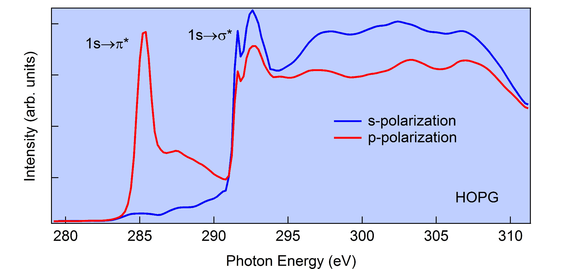
In figure 1 the polarization dependence of C K-edge measured in TEY on HOPG is reported. Depending on the orientation of the electric field vector of the X-rays one can probe predominantly transitions from C-1s to σ*-states (orbitals lying within the graphite plane) or C-1s to π*-states (orbitals normal to the graphite plane).
WARNING: Access to DESY temporarily not available, but the technique is available at the other sites
XAS, XMCD - BACH Beamline @ Elettra Synchrotron
X-ray absorption spectroscopy with selectable polarization, x-ray linear/circular dichroism, time-resolved phenomena, out-of-equilibrium phenomena (pump-probe)
Elettra synchrotron, Apple II undulators; variable polarization (horizontal, vertical, circular ±); beam size on the sample 350x350 (HxV, µm2), vertical size can be reduced on request, flux on sample @10 µm slits (best resolution) (ph./s) 2x10¹²-6x10¹⁰
Hamamatsu fast fluorescence detector; photodiode, setup for x-ray absorption transmission measurement, drain current for total electron yield, Scienta R3000 analyzer for partial electron and Auger yield; setup for measurements in static liquid environment
Heating stages (ebeam, direct current, PBN), ion gun (VG), 4 evaporator ports (CF40), gas inlet valve (variable leak valve), diamond file scraper, cleaver, LEED (OciLEED); evaporators for organic molecules; e-beam evaporator (Omicron) for metals (evaporation at low sample temperatures is also possible)
ARPES and XPS are possible in the same chamber; 4 degree-of-freedom manipulator;. x-ray magnetic circular dichroism (max 0.5 T, measurements in remanence); possibility to apply electric fields (measurements in remanence); possibility to superimpose synchrotron beam to laser beam for pump-probe measurements (100 ps resolution)
XMCD, XAS - High Energy APE Beamline @ Elettra Synchrotron
XCMD, XLMD (X-ray linear magnetic dichroism), LMDAD (linear magnetic dichroism in the angular distribution)
Elettra synchrotron, Apple II undulator; variable polarization (horizontal, vertical, circular ±); beam size on sample 500x200 (HxV, µm2), entry slit reduction to 100x100 possible, zone plate optical condenser reduction to 1x3 possible
Omicron EA 125 analyser; drain current Keithley 6514 picoammeter (for XAS/XMCD/XMLD); 10x10 mm silicon photodiode; total yield 20mm channeltron
T range: 30K- 300K (He flow cryostat) for LT stage (while measuring), 300K-700K for HT stage (while measuring); magnetic field: up to 1000 Oe in pulse mode, up to 200 Oe in continuous mode
XRF, XAS - Hard X-ray Micro/Nano-Probe P06 Beamline @ Petra III Synchrotron
XAS, XRF - Micro XAS Beamline @ Swiss Light Source Synchrotron
The microXAS beamline provides X-ray absorption spectroscopy (XAS) and X-ray fluorescence (XRF) experiments requiring high spatial resolution as well as investigations of time-dependent phenomena in the femtosecond time regime
XRF, XAS (transmission, total electron yield) - PHOENIX Beamline @ Swiss Light Source Synchrotron
The PHOENIX (Photons for the Exploration of Nature by Imaging and XAFS) beamline produces a high flux of soft to hard X-rays from an elliptical undulator (APPLE II). Two different endstations at two branches provide the users the necessary tools to perform X-ray microspectroscopic measurements (µ-XAS and µ-XRF)
Elliptical undulator (APPLE II), flux at 3 keV 1 x 10¹¹ph/s/0.1%BW/0.4A , focused spot size on sample
2.5 µm x 2.5 µm
XAS, XRF, XES - Super XAS Beamline @ Swiss Light Source Synchrotron
The SuperXAS beamline includes a large variety of detection systems and sample environments and is thus attractive for researchers from a variety of fields: e.g. material science, catalysis, environmental science, biology, geology and archeology. Techniques that are available at the beamline include time-resolved X-ray absorption fine structure (XAFS) spectroscopy (minute to the millisecond range), X-ray fluorescence (XRF) and X-ray emission spectroscopy (XES)
XAS, EXAFS - Samba Beamline @ Soleil Synchrotron
Time resolved photoemission XPS, ARPES, XAS, XMCD - Tempo Beamline @ Soleil Synchrotron

