XPEEM/SPEM
PhotoEmission Microscopy
Characterisation Installation 5
X-ray PhotoEmission Electron microscopy (XPEEM) is a parallel (full-field), surface-sensitive imaging method using photo-excited electrons, and is therefore related to the photoemission process. The surface sensitivity of the technique arises from the limited inelastic mean-free path of the characteristic photoelectrons in matter. In this technique, X-ray or UV photons shine the sample surface, and an electron optical column (the PEEM) makes, with the emitted photoelectrons, a magnified image of the surface which can be further filtered in energy with an imaging spectrometer or energy filter. Depending on whether secondary electrons or true core-level electrons (both element- and bonding state-specific) are used for microscopy, the image can be characteristic either of the work function anisotropy, or of heterogeneities in elemental distribution and chemical changes at the surface of interest. Besides imaging in real space, reciprocal space microscopy can also be performed by imaging the diffraction plane of the PEEM, which is equivalent to imaging the parallel component of the valence band photoelectron momentum. The so-called kPEEM technique is equivalent to micro-ARPES and now competes it regarding energy resolution, but is much faster since parallel, angular spectroscopic valence band imaging is performed without any sample rotation.
If many PEEM instruments can be found at synchrotron radiation beamlines, most of them use the absorption channel for element-specific microscopy (XANES-PEEM), and are rarely fitted with an effective (i.e, maintaining energy resolution at high lateral resolution) imaging spectrometer to exploit the photoemission channel. Mature, advanced instrumentation is now available with laboratory sources (including X-rays), without significant degradation of the resulting energy and lateral resolutions, and enhancing very much the accessibility of the technique to research teams.
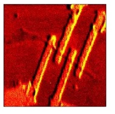
In the Scanning PhotoElectron Microscopy (SPEM) the X-ray photon optics demagnifies the incident photon beam to a small spot onto the sample; an image is acquired by detecting the photoelectron signal while rastering the sample. The microprobe is formed using diffractive optical elements (zone plates); the available spatial resolution is ~ 100 nm. Samples have fixed normal incidence geometry and grazing acceptance angle (60°) of the hemispherical electron analyzer, which enhances the surface sensitivity of the microscope. The SPEM has two operation modes: (i) microspot spectroscopy and (ii) imaging spectromicroscopy. The microspot mode is identical to the XPS spectroscopy, i.e. energy distribution curves are measured from the selected micro-spot area. The best achievable overall energy resolution at room temperature is below 200 meV. The imaging mode maps the lateral distribution of elements by collecting photoelectrons with a selected energy while scanning the specimen with respect to the microprobe.
Research takes place in fields related to surface physics and chemistry, materials science, and nanotechnology. The microscope is a model tool to investigate a wide range of micro and nanostructures, electrochemical reactions, nanocomposite materials and catalytic systems addressing the so called "Material Gap". For instance, the SPEM is an ideal for monitoring in situ dynamic processes, such as mobility of metal on a surface as a function of temperature and/or bias.
The SPEM team has recently developed novel concepts for a new generation of scanning photoelectron microscopes working under more realistic pressure conditions. The first setup, called Dynamic High Pressure (DHP), generates high pressure pulsed gas packets directed to the sample; under the influence of gas pulses samples experience a several tens mbar pressure in a burst instant. The DHP has been already used for the characterisation of electrochemical devices and catalytic materials and is now a standard option for the SPEM. The most recent development is an effusive cell for near-ambient pressure experiments where the highest static pressure achievable is around 0.1 mbar. All these advances are still unique within the photoemission spectromicroscopy community and are paving the way for a new class of in operando experiments where chemical and surface sensitivity together with lateral resolution are needed.
XPEEM, LEEM - SIM Beamline @ Swiss Light Source Synchrotron
The permanent endstation of the SIM (Surfaces/Interfaces Microscopy) beamline is a Photoemission Electron microscope (PEEM), it allows to image samples using the photoelectric effect with very high spatial resolution
Pure permanent magnet helical undulators
flux 1015 photon/s/0.1%BW/0.4A
focused spot size 30µm x 100µm (V x H)
polarization linear: 0 deg (horizontal) to 90 deg (vertical), circular: right / left
SPEM @ ESCAmicroscopy beamline at Elettra
The experimental apparatus allows to carry out a manifold of experiments, aiming at quantitative and qualitative chemical characterisation of morphologically complex materials including chemical reactions and mass transport processes leading to lateral changes in the composition, morphology and electronic properties of materials, at the submicron scale
Hemispherical electron analyzer SPECS Phoibos 100 equipped with 48 anodes MCP
Energy resolution: 10 mV
Field of view: +/- 10°
Typical collection time: 300ms for energy
Spatial resolution ~ 100 nm
Best achievable overall energy resolution at room temperature below 200 meV
Samples with fixed normal incidence geometry and grazing acceptance angle (60°) of the hemispherical electron analyzer

